
Orson L. Moritz, Ph. D.
Associate Professor, Dept. of Ophthalmology and Visual Science, Michael Smith Scholar, W.K. Stell Scholar, CIHR New Investigator
Biography
Associations
UBC Program in Neuroscience
Research
Research Description
My research focuses on the biochemical and cell biological mechanisms underlying the inherited retinal disorder retinitis pigmentosa. This disorder is associated with death of the retinal rod photoreceptors, which are responsible for dim light sensistivity, and delayed death of cone photoreceptors, which are responsible for colour and bright light sensitivity. The research projects underway in my lab are focused on the mechanisms by which mutations in the rhodopsin gene cause photoreceptor death.
Currently, members of my lab are investigating the early mechanisms by which mutations in the rhodopsin gene cause cell death, with a focus on mutations that cause misfolding of the rhodopsin protein. We have recently established that some rhodopsin mutations can be “rescued” by a mechanism involving binding of 11-cis retinal chromophore.
In addition, we are investigating the late mechanisms by which death of rod photoreceptors eventually causes a secondary death of cone photoreceptors, as well as other long-term consequences of rod cell death on the retina, including retinal remodeling and the possibility of promoting retinal regeneration.
Finally, we are interested in the processes by which rhodopsin and other photoreceptor proteins are transported within rod photoreceptor cells, particularly transport to the rod outer segment organelle, a modified 9+0 sensory cilium that is responsible for transduction of light signals into electrical impulses. It is likely that these transport processes are disrupted in certain types of retinitis pigmentosa. Furthermore, it is likely that these transport processes are relevant in other types of sensory cilia.
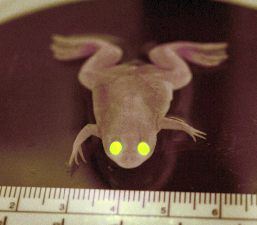
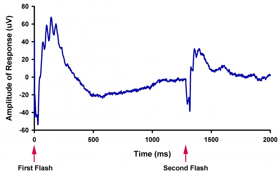
In order to carry out these investigations, my lab employs transgenic Xenopus laevis (frog) models of retinitis pigmentosa. This unique system allows us to generate transgenic animals very rapidly. The X. laevis retina also has excellent properties for our analyses due to the large size of the rod photoreceptors, and the almost equal proportions of rods and cones in the retina. My lab employs many different techniques in our studies, including molecular biology, confocal microscopy, protein chemistry, and electrophysiology.
Recent Publications
Targeting of mouse guanylate cyclase 1 (Gucy2e) to Xenopus laevis rod outer segments. Karan S, Tam BM, Moritz OL, Baehr W.Vision Res. 2011 Sep 12. [Epub ahead of print]
In situ visualization of protein interactions in sensory neurons: glutamic acid-rich roteins (GARPs) play differential roles for photoreceptor outer segment scaffolding. Ritter LM, Khattree N, Tam B, Moritz OL, Schmitz F, Goldberg AF. J Neurosci. 2011 Aug 3;31(31):11231-43.
Recent insights into the mechanisms underlying light-dependent retinal degeneration from X. laevis models of retinitis pigmentosa. Moritz OL, Tam BM. Adv Exp Med Biol. 2010;664:509-15.
The role of rhodopsin glycosylation in protein folding, trafficking, and light-sensitive retinal degeneration. Tam BM, Moritz OL. J Neurosci. 2009 Dec 2;29(48):15145-54.
The dependence of retinal degeneration caused by the rhodopsin P23H mutation on light exposure and vitamin a deprivation.Tam BM, Qazalbash A, Lee HC, Moritz OL. Invest Ophthalmol Vis Sci. 2010 Mar;51(3):1327-34. Epub 2009 Nov 20.
Fourier domain optical coherence tomography as a noninvasive means for in vivo detection of retinal degeneration in Xenopus laevis tadpoles. Lee DC, Xu J, Sarunic MV, Moritz OL. Invest Ophthalmol Vis Sci. 2010 Feb;51(2):1066-70. Epub 2009 Sep 9.